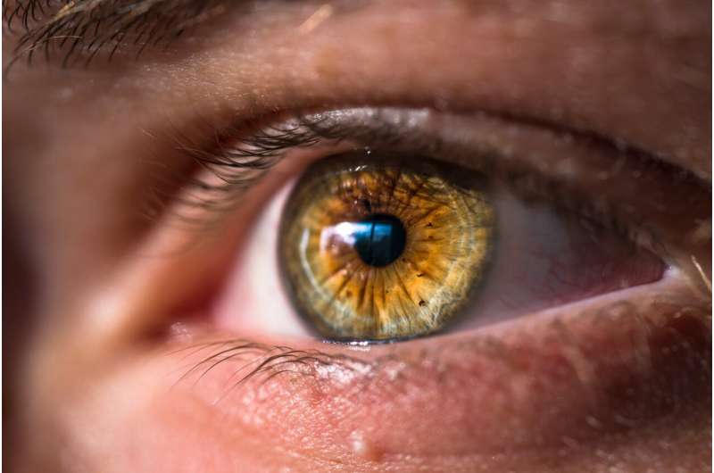
Alterations caused by ALS detected in the retina


Amyotrophic lateral sclerosis (ALS), characterised by harm to brain neurons and to the spinal cord, also affects the tissue of the retina, according to a study headed up by Universidad Complutense de Madrid (UCM).
The research, published in Neural Regeneration Research, indicates that patients with ALS may suffer changes in the microglial cells, responsible for protecting and defending neurons, along with a loss of ganglion cells—the neurons in the retina.
“These alterations detected in this disease can serve as biomarkers for the diagnosis and monitoring of patients with ALS, and to check the efficacy of different treatments through a simple, non-invasive and less expensive form of monitoring”, indicates Pilar Rojas, a researcher at the Ramón Castroviejo Institute for Eye Research (IIORC) at UCM.
As well as describing the changes to the retina, the work also indicates that these modifications evolve over the course of the disease. The microglia change from an M2 state or phenotype (anti-inflammatory) in earlier stages to M1 (proinflammatory) in the final stages.
“This had been observed in the spinal cord of these patients, but not in the retina, which is a step forward, because this may be less invasive to monitor patients”, indicates Rosa de Hoz, from IIORC.
First detection in humans, confirmation in mice
The study has been carried out at the IIORC, in collaboration with the Biochemistry Department of the UCM, the Gregorio Marañón General University Hospital of Madrid, the Ophthalmology Network OFTARED of the Carlos III Health Institute and the RetiBrain Network of the Ministry of Science, Innovation and Universities.
In carrying out the study, the researchers recorded retinal alterations through Optical Coherence Tomography (OCT) – a non-invasive test that performs an analysis of the retina and of the optic nerve, capable of observing minor changes that are often invisible to the human eye”, states the co-author Ana Isabel Ramírez.
Once detected in vivo in humans, they proceeded to confirm this in an animal model through microscopic analysis with immunohistochemical techniques in mice with superoxide dismutase-1 mutation—one of the most common in humans with ALS.
Source: Read Full Article


