An Overview of Caspase Activity

Caspases are proteases: enzymes that catalyze the breakdown of proteins and are part of the cysteine–aspartic acid protease family. They are vital to the mechanism of programmed cell death in animals (apoptosis, pyroptosis, and necroptosis), and also play a fundamental role in inflammation. Due to their involvement in regulating cell death and inflammation, caspase activity is crucial to maintaining homeostasis in the body.
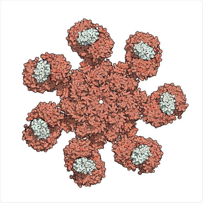
 Image Credit: StudioMolekuul / Shutterstock.com
In mammals, three kinds of caspases exist that have distinct roles. Two of these are involved in apoptosis: initiator caspases are those that initiate apoptosis, while executioner caspases carry out the protein breakdown known as proteolysis. The third group, the inflammatory caspases, are vital to inflammatory cytokine signaling as well as other kinds of cell death.
The significance of research into caspase activity
Recent years have seen a rapid emergence of new information on the molecular mechanisms that control caspase activation. This research is important because it has allowed scientists to attempt to devise new ways to modulate dysfunctional cell death and inflammation pathways that are characteristic of various communicable, inflammatory, malignant, metabolic, and neurodegenerative diseases.
Below, an overview on what has been discovered about caspase activity is given in relation to the two distinct roles of apoptosis and inflammation.
Caspases in apoptosis
Cell death can occur in two ways: necrosis or apoptosis. Cell death that often first comes to mind is that which results due to traumatic injury, poisoning, an infection, or a circumstance that cuts the oxygen supply to the cells. This is referred to as necrosis.
Apoptosis, in contrast, is a necessary part of the life cycle of a cell. It is programmed cell death that is caused by the controlled dismantling of intracellular components in a process that protects surrounding cells from damage, as it impacts only the cells intended to go through cell death.
The process of apoptosis is instituted by the initiator caspases -2, -8, and -9, with -9 being activated on the apoptosome complex to act as the essential initiator for apoptosis signaling through the mitochondrial pathway. Importantly, studies have uncovered that failure to activate caspase-9 is associated with pathophysiological consequences, leading to illness, both mental and physical.
All three initiator caspases become activated at multi-protein activation platforms by homo-dimerization of the monomeric zymogens to induce their role in apoptosis. The long prodomains (>90 amino acids) that are embodied in the initiator caspases make it possible for them to communicate with specific signaling complexes, and bring about the execution stage of apoptosis.
The executioner caspases carry out mass proteolysis, which is the controlled process of protein breakdown into smaller polypeptides or amino acids, resulting in cell death. Research has shown that caspases -3, -6, and -7 are responsible for regulating the execution phase of apoptosis by cleaving multiple structural and repair proteins.
Following activation of a single executioner caspase, further executioner caspases can be cleaved and activated by this initial caspase. This results in accelerated activation of executioner caspases, leading to efficient cell death.
Caspases in inflammation
Caspase-1, -4, -5, and -12 mediate the immune response in humans. The inflammatory caspases are produced in the same way as proapoptotic caspases, as inactive procaspases in resting cells. Engagement with pattern-recognition receptors resulting in the formation of the inflammasome stimulates the activation of the inflammatory caspases.
The inflammasome is established through the processes of leucine-rich repeat-containing (NLR) proteins. The proteins that are held within inactive cells can become stimulated by either an internal or external incident, which initiates their assembly. Once assembled, the NLR monomers bind to the adapter protein apoptosis-associated speck-like protein.
An inactive inflammatory procaspase is recruited into the inflammasome, and this process induces proximity autocatalysis that has the effect of cleaving and activating caspase-1, which then activates the recreation of the proinflammatory cytokines. The immune response is able to behave in a tailor-made way in response to different stimuli because different NLR-driven inflammasomes respond to different stimuli.
There is some evidence to suggest that caspase-12 plays the role of having an anti-inflammatory response through its inhibitory effect on caspase-1; however, the precise mechanisms still remain elusive.
There is still work to be done in order to fully understand the activation of inflammasomes in response to specific situations in the body. An increased understanding could lead to a clearer view of immune system diseases and potentially result in the development of new treatments that appropriate the immune response.
Caspases and illness
Tumorigenesis is unsurprisingly supported by malfunction of the caspases, as they play a vital role in cell death, a process that can act as a preventative measure of cancer by killing faulty cells before they become oncogenic. Cancer is not the only disease that the caspases have been found to be involved in.
The role of the caspases has been implicated in the development and maintenance of numerous illnesses, including cancer, inflammatory diseases, gout, cryopyrin-associated periodic syndromes, type 2 diabetes, familial Mediterranean fever, pyogenic sterile arthritis, pyoderma gangrenosum, acne syndrome, hyperimmunoglobulinemia D with periodic fever syndrome, systemic-onset juvenile idiopathic arthritis, Alzheimer’s disease, Kawasaki disease, and autoimmune lymphoproliferative syndrome.
Further exploration into the mechanisms of caspase activity has the potential to impact the diagnosis, treatment, prognosis, and prevention of these illnesses.
Sources:
- Kumar, S. (2006). Caspase function in programmed cell death. Cell Death & Differentiation, 14(1), pp.32-43. https://www.nature.com/articles/4402060
- McIlwain, D., Berger, T., and Mak, T. (2013). Caspase Functions in Cell Death and Disease. Cold Spring Harbor Perspectives in Biology, 5(4), pp.a008656-a008656. https://cshperspectives.cshlp.org/content/5/4/a008656.full
- Shalini, S., Dorstyn, L., Dawar, S., and Kumar, S. (2014). Old, new, and emerging functions of caspases. Cell Death & Differentiation, 22(4), pp.526-539. https://www.nature.com/articles/cdd2014216
- Van Opdenbosch, N., and Lamkanfi, M. (2019). Caspases in Cell Death, Inflammation, and Disease. Immunity, 50(6), pp.1352-1364. https://www.ncbi.nlm.nih.gov/pubmed/31216460
Further Reading
- All Cell Death Content
- What is Ferroptosis?
- How to Analyze Cellular Necroptosis
- Pyroptosis Pathway and Triggers
- Avoiding Cell Death in Fluorescence Microscopy
Last Updated: Dec 11, 2019

Written by
Sarah Moore
After studying Psychology and then Neuroscience, Sarah quickly found her enjoyment for researching and writing research papers; turning to a passion to connect ideas with people through writing.
Source: Read Full Article


