Study links early life adversity, microglia dysfunction, to aberrant adult stress responses, mental illness
. DOI: 10.1016/j.celrep.2022.110600")
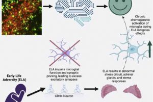
The poor function of microglia, the brain’s immune cells in individuals exposed to early life adversity (ELA) promotes aberrant responses to stress in adulthood that may be linked to mental illness, according to a new study led by researchers at the University of California, Irvine. During brain development, microglia prune unnecessary synapses resulting in the formation of refined, functional circuits. Disruption of that process leaves too many synapses, changing the behavioral and hormonal responses to further stresses later in life.
The study, titled “Early-stress induced impairment of microglial pruning of excitatory synapses on immature CRH-expressing neurons provokes aberrant adult stress responses,” was published online today in Cell Reports.
“Much of neuroscience and study of brain diseases has focused on the brain’s neurons. This study highlights that in addition to neurons, other brain cells, and especially immune cells, play crucial roles in brain health and disease,” said Tallie Z. Baram, distinguished professor in the Departments of Anatomy & Neurobiology, Pediatrics, Neurology, and Physiology & Biophysics at the UCI School of Medicine. “Neuroimmune interactions are a novel, important avenue to understanding and treating several brain disorders and mental illness and have been linked by other UCI researchers to Alzheimer’s disease.”
Brain development is governed by both genetics and early life experiences. Several mental illnesses, characterized by aberrant reactions to stress, often arise after ELA but its effect on stress-related brain circuit maturation was unclear. In the study, led by UCI Postdoctoral Fellow Jessica Bolton, who is now an Assistant Professor at Georgia State University, transgenic mice of both sexes were housed in temperature-controlled, quiet, uncrowded conditions on a schedule of 12 hours of light and 12 hours of dark, with free access to food and water. The control dams were place in cages with standard amounts of corn husk bedding and nesting material. The ELA dams received only one-half nestlet and reduced bedding materials.
Researchers found that ELA increased functional excitatory synapses onto stress-sensitive hypothalamic corticotropin-releasing hormone-(CRH) expressing neurons, resulting from disrupted developmental synapse pruning by adjacent microglia in the mouse pups. These neuroimmune interactions during development thus made the CRH cells, and they mice, more vulnerability to stress.
Source: Read Full Article


