A Guide to Fluorescence

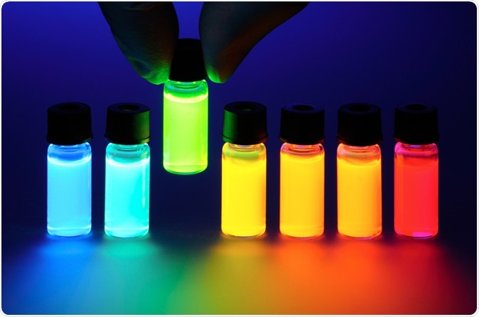
The phenomenon of fluorescence is a form of luminescence, and it is exploited by techniques that have innovated ways to utilize its light emittance to gain insights into target materials or substances.

Image Credit: Denis Larkin/Shutterstock.com
Fluorescence techniques were designed to observe and analyze the composition of samples, as well as interactions between various biomolecules within living cells, including proteins, nucleic acids, or lipids. Numerous biophysical methods exist that take advantage of fluorescence.
Fluorophores that emit light are used to identify the presence of various molecules within a bio-molecular structure. Quite often, fluorescence techniques are used to investigate the contents of live cells.
How the fluorescence process works
The essence of how the fluorescence process works lies within the ability of the fluorophore to emit light after it has been exposed to particular wavelengths of light during an absorption phase. The fluorophores are selected to respond to certain stimuli within a biological sample. The process works in a three-stage process:
Excitation
Excitation is the first stage of the process. This is where energy is supplied to the fluorophore in the form of a light source, which could be an incandescent lamp or a laser. Incident photons have the impact of exciting the fluorophore after it has been absorbed, initiating an excited electronic singlet state.
Excited-State Lifetime
After the excitation phase, the molecules remain in the excited state for a length of time, usually between 1 and 10 nanoseconds. Within the excitation phase, the fluorophore progresses through conformational changes in addition to interacting with other molecules in its environment. The impact of these events is important.
Firstly, the fluorescent emission is initiated by the dissipation of the energy that was passed to the fluorophore during excitation. Also, not all the molecules that became excited, return to their normal state. These processes can be analyzed and are important for identifying the components and interactions within a substance.
Fluorescence Emission
After the initial excitation-state phase, energy dissipates from the molecules, and the fluorophore returns to its ground state. This release of energy produces the fluorescent emission, and it can be observed as a longer wavelength than was presented at the time of excitation because the photon has lost some energy.
The length of time that it takes for the fluorophore to emit its energy depends on the interactions it made with its environment, and this can tell researchers about the sample it has been added to. The difference observed between the initial wavelength and the wavelength emitted is called the Stokes shift.
The fluorophore can be used multiple times, excited over again and detected for analysis. Usually, the fluorophore can be used in numerous tests, however, sometimes is becomes irreversibly damaged in a process called photobleaching. Photobleaching occurs during the excited state, due to cleaving covalent bonds or through non-specific reactions with molecules in the environment. Because under normal circumstances a single fluorophore can be used to generate thousands of photons is what makes it a highly sensitive detection technique.
The emission of light during fluorescence is always different from the excitation wavelength because of the loss of excitation energy. Also, the intensity of the emission intensity is directly related to the amplitude of the fluorescence excitation spectrum during the phase.
Fluorescence detection
There are four key parts common to most fluorescence techniques. As has been highlighted, there first needs to be a source of excitation (a light source), a fluorophore, wavelength filters, which are required for the identification of emission photons against excitation photons, and a form of detector that can pick up the emission photons, usually recording them in the form of an electronic signal.
The fourth and final factor essential to fluorescence techniques, that of a detector, exists in four types. The first is spectrofluorometers and microplate readers, these detectors are used to measure the average properties of bulk samples.
Fluorescence microscopes are the second kind, and they present fluorescence as a function of spatial coordinates. This can be in either two or three dimensions, and the technique is usually used for microscopic samples.
The third kind is the fluorescence scanner, like microarray readers, for example. These detectors also present fluorescence as a function of spatial coordinates, but only in two dimensions. This technique is used for macroscopic objects, like electrophoresis gels and chromatograms, for instance.
The final kind of detector is the flow cytometer, and it measures fluorescence per cell in a flowing stream.
Fluorescence output of fluorophores
Different fluorophores are available for use in fluorescence techniques. Organic dye and autofluorescent protein fluorophores are the two main types of dyes used.
The fluorescent quality of the fluorophore is reliant on factors such as how capable it is of undergoing repeated excitation/emission cycles, and how efficient it is at absorbing and releasing photons.
The fluorescent signals of a fluorophore can be enhanced by simply increasing how many fluorophores are available for detection in a sample. Signals can also be enhanced by using an avidin-biotin or antibody–hapten secondary detection techniques, or by using enzyme-labeled secondary detection reagents alongside fluorogenic substrates. Finally, the signal can also be enhanced by using probes that contain multiple fluorophores.
Sources:
Combs, C. (2010). Fluorescence Microscopy: A Concise Guide to Current Imaging Methods. Current Protocols in Neuroscience. https://www.ncbi.nlm.nih.gov/pmc/articles/PMC3805368/
Combs, C. and Shroff, H. (2017). Fluorescence Microscopy: A Concise Guide to Current Imaging Methods. Current Protocols in Neuroscience, pp.2.1.1-2.1.25. currentprotocols.onlinelibrary.wiley.com/doi/abs/10.1002/cpns.29
(2009) Fluorescence Detection Techniques. In: Demchenko A.P. (eds) Introduction to Fluorescence Sensing. Springer, Dordrecht https://link.springer.com/chapter/10.1007/978-1-4020-9003-5_3#citeas
Schermelleh, L., Heintzmann, R. and Leonhardt, H. (2010). A guide to super-resolution fluorescence microscopy. The Journal of Cell Biology, 190(2), pp.165-175. http://jcb.rupress.org/content/190/2/165.short
Further Reading
- All Fluorescence Content
- What is Fluorescence Spectroscopy?
- How do Epifluorescence Microscopes Work?
- GFP-tagging in Fluorescence Microscopy
- Fluorescence Quenching
Last Updated: Feb 3, 2020

Written by
Sarah Moore
After studying Psychology and then Neuroscience, Sarah quickly found her enjoyment for researching and writing research papers; turning to a passion to connect ideas with people through writing.
Source: Read Full Article


